|
Okay, so last semester didn’t actually involve any tears, but I spent a lot of time looking at salamander blood smears under a microscope, and it wasn’t always easy. The majority of time I spend in lab was dedicated to counting the different cells that I saw. As I peered through the microscope, I identified different types of white blood cells. By counting these cells and looking at differences among the ratios of different cell types we can estimate how stressed a salamander is. However, identifying cells under a microscope after learning what they look like from pictures takes some getting used to. It is complicated to learn how to identify cells under a microscope from a reference pictures because the cells under the microscope don’t necessarily look the same as in a picture. Intracellular variation can be high! Therefore, there is a lot of trial and error in learning to identify cells under a microscope until you get the hang of it. Practice, practice, practice!  The types of cells that I identified in the blood smear included erythrocytes (red blood cells) and leukocytes (white blood cells). When looking at slide, the erythrocytes are abundant and easy to identify. It is interesting that they are nucleated in amphibians, which is different than our own red blood cells. Since erythrocytes are easy to spot, the catch is not finding them, but counting them as there can be dozens in one field of view!
To identify a lymphocyte I looked for a round agranular cell with the nucleus taking up more of the cytoplasm than an erythrocyte. Neutrophils have multi-lobed nuclei and were easy to spot since they stood out from all the other cells with their amorphous nuclei. Eosinophils had a slightly pinkish color to the cytoplasm when being observed under the microscope and were a little more granulated but also had a lobed nuclei. Basophils are very rare to spot, but they are very dark and granulated. I was able to find monocytes by looking for a nucleus that was less rounded and sometimes had a dimple in it making it appear like a kidney bean (although not always). 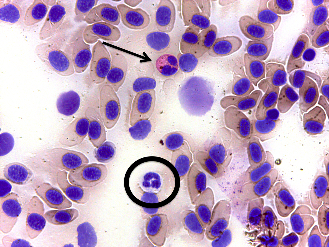 The picture above is from one of the slides that I counted. The arrow is pointing to an eosinophil and the circle is around a neutrophil. It's not just these easy to identify cells that are on the slides! When counting the slides you occasionally come across thrombocytes, cells that were crushed, or dividing cells. I was careful not to accidentally include them in my count, by mistaking them for something else. I don't count every single cell that I see, only the ones that I am supposed to identify. Some of other cells visible are not relevant to the counts I do.  Taking a look at the picture below you can see some of the challenges I encountered with the slides. Just look at that mess! Not every field of view is countable — Imagine trying to figure out how many cells are on that field of view, let alone distinguishing what they are. Spoiler alert, it's not going to happen, but that's ok. I just skipped over these views and moved to more countable ones. When I first started counting slides I thought that I was never going to get the hang of it as it seemed overwhelming and confusing! Luckily, the more time I spent doing it the easier and more natural it became. By the end of the semester I was able to count the slides much faster and with much more confidence!  ~Alexa DeMaio
0 Comments
|
The Slime Times is written by the Stress After Dark Creative Inquiry TeamScience isn't all white lab coats and ivory pillars, sometimes you just need to get slimy. Archives
August 2016
Categories |


 RSS Feed
